Basic Technique of Plant Tissue Culture
Basic Technique of Plant Tissue Culture
Introduction
Plant tissue culture, the growth of plant cells outside an intact plant, is a technique essential in many areas of the plant sciences. Cultures of individual or groups of plant cells, and whole organs, contribute to understanding both fundamental and applied science.
The application of modern biotechnology techniques like plant tissue culture has proven to be a powerful tool in medicinal plant improvement programs. Scientists hit upon a technique where by not only can these plants be preserved from being lost but are also able to develop a complete plant from a small plant part.
Basically plant tissue culture is the technique of growing plant cells, tissues and organs in an artificial, prepared nutrient medium, static or liquid, under aseptic conditions. These explants divide and gradually develop into an unorganized mass of cells called “callus” and subsequently differentiate to form plant or directly give rise to shoots or embryos.
Plant tissue culture is now the major component of technologies which are applied in plant biotechnology. Advances made in genetic engineering and molecular biology can be made manifest in plant through the application of various techniques developed in the field of plant tissue culture. Many of the crop plants regarded as recalcitrant are now amenable to regeneration in vitro using cultured protoplasts, cells or calli, thus each of them can be used as a tool in plant genetic manipulation programs. Considerable progress has been made with regard to the development of media or techniques as well as in understanding the basic aspects, such as cell culture, cellular totipotency. Development of plant tissue culture is closely linked to improvement of techniques of protoplast, cell, tissue and organ culture followed by the success achieved in regenerating whole plants from culture plant materials.
Knowledge of plant tissue culture has contributed greatly to our understanding of the factors responsible for growth, metabolism, differentiation and morphogenesis of plant cells. Plant tissue culture is presently of great interest to molecular biologists, plant breeders and industrialists. Tissue culture methods have been used as a tool for the propagation of genetically manipulated superior clones and for ex-situ conservation of valuable germplasm. The progress in use of cell or tissue culture in producing pathogen free plant as well as in the synthesis of many important secondary compounds (including pharmaceuticals) has been very significant.
Though a considerable progress has been made in tissue culture of plant species, the method is not widely applicable in its present state for cloning, improvement, somaclonal variation, disease resistance, protoplast culture and genetic engineering of many endangered medicinal plant, therefore basic information generated will be useful on these lines of work for specific and selected cases for developing clones for fodder, medicine and various types of resistance.
Most methods of plant transformation applied to GM crops require that a whole plant is regenerated from isolated plant cells or tissue which have been genetically transformed. This regeneration is conducted in vitro so that the environment and growth medium can be manipulated to ensure a high frequency of regeneration. In addition to a high frequency of regeneration, the regenerable cells must be accessible to gene transfer by whatever technique is chosen. The primary aim is therefore to produce, as easily and as quickly as possible, a large number of regenerable cells that are accessible to gene transfer. The subsequent regeneration step is often the most difficult step in plant transformation studies. However, it is important to remember that a high frequency of regeneration does not necessarily correlate with high transformation efficiency. This chapter will consider some basic issues concerned with plant tissue culture in vitro, particularly as applied to plant transformation. It will also look at the basic culture types used for plant transformation and cover some of the techniques that can be used to regenerate whole transformed plants from transformed cells or tissue. Practically any plant transformation experiment relies at some point on tissue culture. There are some exceptions to this generalization, but the ability to regenerate plants from isolated cells or tissues invitro underpins most plant transformation systems.
Plasticity and Totipotency
Two concepts, plasticity and totipotency, are central to understanding plant cell culture and regeneration. Plants, due to their sessile nature and long life span, have developed a greater ability to endure extreme conditions and predation than have animals. This plasticity allows plants to alter their metabolism, growth and development to best suit their environment. Particularly important aspects of this adaptation, as far as plant tissue culture and regeneration are concerned, are the abilities to initiate cell division from almost any tissue of the plant and to regenerate lost organs or undergo different developmental pathways in response to particular stimuli. When plant cells and tissues are cultured in vitro they generally exhibit a very high degree of plasticity, which allows one type of tissue or organ to be initiated from another type. In this way, whole plants can be subsequently regenerated.
This regeneration of whole organisms depends upon the concept that all plant cells can, given the correct stimuli, express the total genetic potential of the parent plant. This maintenance of genetic potential is called ‘totipotency’. Plant cell culture and regeneration do, in fact, provide the most compelling evidence for totipotency.
Types of Tissue Culture
Cultures are generally initiated from sterile pieces of a whole plant. These pieces are termed ‘explants’, and may consist of pieces of organs, such as leaves or roots, or may be specific cell types, such as pollen or endosperm. Many features of the explant are known to affect the efficiency of culture initiation. Generally, younger, more rapidly growing tissue (or tissue at an early stage of development) is most effective. Several different culture types most commonly used in plant transformation studies will now be examined in more detail.
Callus Culture
Explants, when cultured on the appropriate medium, usually with both an auxin and a cytokinin, can give rise to an unorganised, growing and dividing mass of cells. It is thought that any plant tissue can be used as an explant, if the correct conditions are found. In culture, this proliferation can be maintained more or less indefinitely, provided that the callus is subcultured on to fresh medium periodically. During callus formation there is some degree of dedifferentiation (i.e. the changes that occur during development and specialization are, to some extent, reversed), both in morphology (callus is usually composed of unspecialised parenchyma cells) and metabolism. One major consequence of this dedifferentiation is that most plant cultures lose the ability to photosynthesise. This has important consequences for the culture of callus tissue, as the metabolic profile will probably not match that of the donor plant. Callus culture is often performed in the dark (the lack of photosynthetic capability being no drawback) as light can encourage differentiation of the callus.
Cell-suspension Cultures
Callus cultures, broadly speaking, fall into one of two categories: compact or friable. In compact callus the cells are densely aggregated, whereas in friable callus the cells are only loosely associated with each other and the callus becomes soft and breaks apart easily. Friable callus provides the inoculum to form cell-suspension cultures. Explants from some plant species or particular cell types tend not to form friable callus, making cell-suspension initiation a difficult task. The friability of callus can sometimes be improved by manipulating the medium components or by repeated subculturing. The friability of the callus can also sometimes be improved by culturing it on ‘semi-solid’ medium (medium with a low concentration of gelling agent). When friable callus is placed into a liquid medium (usually the same composition as the solid medium used for the callus culture) and then agitated, single cells and/or small clumps of cells are released into the medium.
Protoplasts Culture
Protoplasts are plant cells with the cell wall removed. Protoplasts are most commonly isolated from either leaf mesophyll cells or cell suspensions, although other sources can be used to advantage. Two general approaches to removing the cell wall (a difficult task without damaging the protoplast) can be taken—mechanical or enzymatic isolation. Mechanical isolation, although possible, often results in low yields, poor quality and poor performance in culture due to substances released from damaged cells.
Enzymatic isolation is usually carried out in a simple salt solution with a high osmoticum, plus the cell wall grading enzymes. It is usual to use a mix of both cellulase and pectinase enzymes, which must be of high quality and purity.
Protoplasts are fragile and easily damaged, and therefore must be cultured carefully. Liquid medium is not agitated and a high osmotic potential is maintained, at least in the initial stages. The liquid medium must be shallow enough to allow aeration in the absence of agitation. Protoplasts can be plated out on to solid medium and callus produced. Whole plants can be regenerated by organogenesis or somatic embryogenesis from this callus. Protoplasts are ideal targets for transformation by a variety of means.
Root Cultures
Root cultures can be established in vitro from explants of the root tip of either primary or lateral roots and can be cultured on fairly simple media. The growth of roots in vitro is potentially unlimited, as roots are indeterminate organs. Although the establishment of root cultures was one of the first achievements of modern plant tissue culture, they are not widely used in plant transformation studies.
Shoot Tip and Meristem Culture
The tips of shoots (which contain the shoot apical meristem) can be cultured in vitro, producing clumps of shoots from either axillary or adventitious buds. This method can be used for clonal propagation. Shoot meristem cultures are potential alternatives to the more commonly used methods for cereal regeneration (see the Case study below) as they are less genotype-dependent and more efficient (seedlings can be used as donor material).
Embryo Culture
Embryos can be used as explants to generate callus cultures or somatic embryos. Both immature and mature embryos can be used as explants. Immature, embryo-derived embryogenic callus is the most popular method of monocot plant regeneration.
Microspore Culture
Haploid tissue can be cultured in vitro by using pollen or anthers as an explant. Pollen contains the male gametophyte, which is termed the ‘microspore’. Both callus and embryos can be produced from pollen. Two main approaches can be taken to produce in vitro cultures from haploid tissue. The first method depends on using the anther as the explant. Anthers (somatic tissue that surrounds and contains the pollen) can be cultured on solid medium (agar should not be used to solidify the medium as it contains inhibitory substances). Pollen-derived embryos are subsequently produced via dehiscence of the mature anthers. The dehiscence of the anther depends both on its isolation at the correct stage and on the correct culture conditions. Anthers can also be cultured in liquid medium, and pollen released from the anthers can be induced to form embryos, although the efficiency of plant regeneration is often very low. Immature pollen can also be extracted from developing anthers and cultured directly, although this is a very time-consuming process.
Plant Regeneration
Having looked at the main types of plant culture that can be established in vitro, we can now look at how whole plants can be regenerated from these cultures. In broad terms, two methods of plant regeneration are widely used in plant transformation studies, i.e. somatic embryogenesis and organogenesis.
Somatic Embryogenesis
In somatic (asexual) embryogenesis, embryo-like structures, which can develop into whole plants in a way analogous to zygotic embryos, are formed from somatic tissues (Figure 2.2). These somatic embryos can be produced either directly or indirectly. In direct somatic embryogenesis, the embryo is formed directly from a cell or small group of cells without the production of an intervening callus. Though common from some tissues (usually reproductive tissues such as the nucleus, styles or pollen), direct somatic embryogenesis is generally rare in comparison with indirect somatic embryogenesis. In indirect somatic embryogenesis, callus is first produced from the explant. Embryos can then be produced from the callus tissue or from a cell suspension produced from that callus.
Organogrnesis
Somatic embryogenesis relies on plant regeneration through a process analogous to zygotic embryo germination. Organogenesis relies on the production of organs, either directly from an explant or from a callus culture. There are three methods of plant regeneration via organogenesis. The first two methods depend on adventitious organs arising either from a callus culture or directly from an explants . Alternatively, axillaries bud formation and growth can also be used to regenerate whole plants from some types of tissue culture.
Benefits of Tissue Culture
- Tissue culture offers numerous significant benefits over traditional propagation methods:
- Much faster rates of growth can be induced in vitro than by traditional means.
- It may be possible to multiply in vitro plants that are very difficult to propagate by cuttings or other traditional methods.
- Large numbers of genetically identical clones may be produced.
- Seeds can be germinated with no risk of damping off/predation.
- Under certain conditions, plant material can be stored in vitro for considerable periods of time with little or no maintenance.
- Tissue culture techniques are used for virus eradication, genetic manipulation, somatic hybridization and other procedures that benefit propagation, plant improvement, and basic research.
Application of Tissue Culture
- Micropropagation: Rapid vegetative multiplication of valuable plant material for agriculture, horticulture, and forestry.
- Production of disease-free plants: When the apex of shoot is used for multiplication by tissue culture, we get disease free plants because the shoot apical meristem, a group of dividing cells at the tip of a stem or root, is free from pathogens.
- Plant Breeding: Tissue culture has also been successfully used in plant breeding programmes.
- Production of disease and pest-resistant plants: Plants grown from tissue culture usually pass trough callus phase and show many variations. These show some agronomic characteristics like tolerance to pests, diseases, etc.
- Cloning: Genetically identical plants derived from an individual are called clones. Processes that produce clones can be put under the term ‘cloning’. This includes all the methods of vegetative propagation such as cutting, layering, and grafting. Propagation by tissue culture also helps in producing clones. Using the shoot tip, it is possible to obtain a large number of plantlets. This technique is used extensively in the commercial field for micropropagation of ornamental plants like chrysanthemum, gladiolus, etc. and also crops such as sugar cane, tapioca, and potato. Thus an unlimited number of plants that are genetically similar or are clones can be produced in a short span of time by tissue culture.
- Large-scale propagation: To bridge the gap between research and application, the Department of Biotechnology, Government of India sponsored the setting-up of two pilot-scale facilities for large-scale propagation of elite planting material of forest trees through tissue culture. One of these facilities has been established at TERI’s 36-hectare-campus in Gual Pahari, Haryana with an annual capacity of a million plantlets. Research at these facilities focuses exclusively on developing new protocols for mass cloning of elite planting material, mainly of trees.
Over 4 million plants have been dispatched for field plantation from these facilities. The tissue culture-raised plants are presently being evaluated under field conditions. This is being done in tandem with the forest departments of Haryana, Uttar Pradesh, Madhya Pradesh, Bihar, Jammu and Kashmir, and Orissa. For initial screening for phenotypically superior trees, only a few hundred plantlets of the same are raised and tested under various agroclimatic zones. The best clones are then mass multiplied and monitored regularly for their performance. Field data suggest a more than 90% survival rate even in the harsh conditions of Aravalis without the life-saving irrigation.
History of Plant Tissue Culture
This cell is evidently the repository of all the information necessary for its subsequent growth into a multicellular, highly organized, complex but co-ordinated system. This tiny totipotent cell conceals the potential for differentiation. The differentiated somatic cells in a plant carry out specialized activities and appear to have surrendered their totipotency in the bargain. The idea of totipotency of plant cells was put forward by G. Haberlandt, the great German physiologist who in 1902 suggested that “one could successfully cultivate artificial embryos from vegetative cells”. He isolated cells from a number of higher plants and maintained them alive in a viable state in simple nutrient solutions for about 10 days. During this period, cell swelling and wall thickening occurred, but the cells failed to divide. Haberlandt’s attempt to grow vegetative cells in an artificial medium did not succeed due to lack of proper techniques and unfortunate choice of highly specialized materials but it opened up new vistas in morphogenesis.
This cell is evidently the repository of all the information necessary for its subsequent growth into a multicellular, highly organized, complex but co-ordinated system. This tiny totipotent cell conceals the potential for differentiation. The differentiated somatic cells in a plant carry out specialized activities and appear to have surrendered their totipotency in the bargain. The idea of totipotency of plant cells was put forward by G. Haberlandt, the great German physiologist who in 1902 suggested that “one could successfully cultivate artificial embryos from vegetative cells”. He isolated cells from a number of higher plants and maintained them alive in a viable state in simple nutrient solutions for about 10 days. During this period, cell swelling and wall thickening occurred, but the cells failed to divide. Haberlandt’s attempt to grow vegetative cells in an artificial medium did not succeed due to lack of proper techniques and unfortunate choice of highly specialized materials but it opened up new vistas in morphogenesis.
- Schleiden M. J., (1838) suggested totipotentiality of cells. Vochtung in 1878 obderved that cells along a stems length were capable of generating roots or shoots. Sachs J., (1882) synthesizes organ-forming substances that are polarly distributed.
- Haberlandt G., Sitzungsber Akad. Wiss. Wien,(1902) attempt tissue culture using monocots for first time but unsuccessful. A German botanist was the first to generate tissue from fully differentiated tissue.
- Hannig B., (1904) first attempt in embryo culture of selected Crucifers. Laibach successfully reared embryos that were otherwise unviable using tissue culture. Several new hybrids have since 'evolved’ using tissue culture that would otherwise have been unviable at the embryo stage.
- Kuster E. (1909) attempt fusion of plant protoplasts though the products failed to survive.
- Molliard M., C. R. (1921) cultivation of fragments of plant embryos.
- Knudson L., (1922) report asymbiotic germination of orchid seeds.
- Robbins W. J., (1922) report success In vitro culture of root tips. Blumenthal F. and Meyer P. Z. Krebsforsch (1924) present callus formation on carrot root explants by use of lactic acid. Laibach F., (1925) work on embryo culture for interspecific crosses in Linum spp.Knudson L.,(1925) work on symbiotic germination of orchid seeds.
- Laibach F., J Hered., (1929) worked on embryo culture to avoid cross incompatibility in Linum spp.
- Guatheret R. J., C. R. (1934) attempt in vitro culture of cambial tissues of different trees and shrubs which failed.
- White P. R., (1934) successfully reared tomato root cultures using a medium containing three B vitamins; Pyrodoxine, thiamine and Nicotinic acid, also inorganic salts and sucrose.
- Kogl F. et al., (1934) identified the first plant hormone, IAA, leading to cell enlargement.
- LaRue C. R., (1936) worked on embryo culture of different gymnosperms.
- Gautheret R. J., C. R., Nobecourt P., White P. R., (1939) Successfully worked on continuously growing cambial cultures of carrot and tobacco.
- Gautheret R. J., (1940) Culture of cambial tissue of Ulmus to study adventitious shoot formation. Overbeek J. van et al., (1941) used Coconut Milk for growth and development of very young Datura embryos.
- Gautheret R. (1942) Observed secondary metabolites in plant callus cultures.Braun A. C. (1943-1950) work on tumor-inducing principle of crown gall tumors identified.
- Skoog F., (1944) Firstly work on In vitro culture of tobacco used to study adventitious shoot formation.
- Loo S. W., (1945) Cultivated of excised stem tips of Asparagus.Ball Skoog F. and Tsui C., (1948) Formation of adventitious shoots and roots in tobacco.
- Ball E., (1950) work on Organs regenerated from callus of Sequoia. Morel G. C. R. Acad., (1950) First successful cultures of Monocots using coconut milk.
- Nitsch J. P., (1951) Culture of excised ovaries In vitro. Skoog F., (1951) Chemical control of growth and organ formation in culture was demonstrated.
- Morel G. and Martin C., (1952) Virus-free Dahlia through meristem culture. They for the first time obtained virus free dahlia plants from diseased individuals by cutting healthy shoot tips. Morel G. and Martin C., (1952) First successful micro-grafts.
- Miller C. et al., (1955) discovery, structure and synthesis of Kinetin. Reinert J. and White P. R., (1956) In vitro cultivation of normal and tumor tissues of Picea glauca.
- Production of substances from plant tissue culture of Phaseolus by Routien J. B. and Nickell L. G. (1956)
- Skoog F. and Miller C. O., (1957) discovery that root or shoot formation in culture depends on auxin:cytokinin ratio.
- Maheshwari P. and Rangaswamy N. S., (1958) regenerate somatic embryos from nucellus of Citrus ovules. Reinert J. and Steward F. C., (1958) work on pro-embryo formation in callus clumps and cell suspension of carrot. Steward F. C. et al., (1958) growth and development the explant in suspension cultures.
- Tulecke W. and Nickell L. G. (1959) produced large amounts (134 L) of plant tissue by submerged culture.
- Guha S. and Maheshwari S. C. (1964) produced first haploid plants from Datura androgenesis. Haploid plants from pollen grains were first produced by Maheshwari and Guha, when they cultured anthers of Dhatura.
- Pierik R. L. M.,(1967) worked on flower induction in Lunaria annua by vernalization In vitro. Kaul B. and Staba E. J., (1967) yields secondary products in cell culture equal to those of intact plants of Ammi visnaga. Bourgin J. P. and Nitsch J. P., (1967) work on Haploid plants from pollen grains of tobacco.
- Ericksson T. and Jonassen K., (1969) isolated protoplast from suspension culture of Hapopappus gracilis. Carlson P. S. (1970) selected biochemical mutants in tobacco. Kasha K. J. and Kao K. N., (1970) work on Hybrid embryo culture and subsequent chromosome elimination for haploid production in Barley. Power J. B. et al., (1970) work on Protoplast fusion.
- Seibert M., (1976) Shoot induction from cryo-preserved shoot tips of carnation.
- Power J. B. et al., (1976) Protoplast fusion of Petunia hybrida with P. parodii.
- Bomhoff G. et al., (1976) Octopine and Nopaline synthesis and break-down is regulated by Ti plasmid.
- Larkin P. J. and Scowcroft W. R., (1981) Introduction of the term somaclonal variaion.
- Sidorov V. et al, (1981) Isolation of auxotrophs by cell colony screening in haploid protolasts of Nicotiana plumbaginifolia treated with mutagens. Shuler M. L., Ann. (1981) Use of a hollow fiber reactor for secondary metabolite production.
- Pelletier G. et al., (1983) intergeneric cybrid in radish and rape. First industrial production of secondary metabolites by suspension cultures of Lithospermum spp. by Mitsui Petrochemicals (1983).
- Wolters B. and Eilert U. Dtsch. (1983) Beneficial use of elicitors in cell suspension cultures. A more recent development in tissue culture is protoplast culture. This technique has allowed the crossing of plants by protoplast fusion, somatic hybrids have thus been produced of Nicotiana glauca x Nicotiana langsdorfii (Bhojwani et al, 1983). Zambryski P. et al., (1983) Co-integrate type of vectors designed for Agrobacterium transformation.
- Kaeppler H. F. et al., (1990) Silicon carbide fiber-mediated DNA delivery in plant cells. Tissue culture technique is a promising option for in vitro multiplication and is gaining acceptance for conservation of medicinal plants (Sharma and Chandel 1992, Fay 1994 and Chandel et al. 1996). Tissue culture of breeding programs and mass clonal production is potent for mass production (Thorpe and Biondi 1982)
Materials and Methods
- A tissue culture laboratory should be equipped with following facilities:
- Sufficient space for washing, sterilization of glassware and other equipments.
- Preparation, sterilization and storage of nutrient medium.
- Aseptic transfer conditions.
- Growing cultures in incubators
- Biochemical analysis of the material microscopic studies of cells and tissues etc.
- Laboratory equipments for Plant Tissue Culture
- Equipments required for the preparation of media:
- Gas, water and electricity supplies
- Different types of glassware
- Hot plate and magnetic stirrer
- Coarse balance (accurate to 0.01 gm.)
- Sensitive balance (accurate to 0.1 mg)
- Micro-wave oven.7. Millipore filters system
- Automatic media dispenser
- pH meter
- Distillation apparatus
- De-Ionizer
- Wide range of chemicals
- Timer for sterilization process
- Metal autoclave racks
- Material for vessel closure
- Autoclave or pressure cooker
- Refrigerator for stock solutions
- Drying and draining racks
- Chemicals and Equipments required for the Preparation and Isolation of Tissue Cultures:
- Laminar airflow cabinet
- Stereomicroscope
- Flaming instrument
- Dry sterilizer
- Filter paper
- Petri dishes
- Low speed centrifuge
- Sodium hypochlorite
- 96% alcohol
- General chemicals and equipment required for Preparation and Isolation of Tissue Cultures
- Drying oven for Glassware
- Automatic dishwasher
- Cleaning materials
- Detergent
- Trolley
- Small deep - freeze
- Chromic acid bath
- Equipment required for the Tissue culture storage room
- Temperature control (17-200C)
- Lightning
- Shelving for culture rocks
- Timer for day-length control
- Shaker
- For the growth of experimental material and further growth of in vitro-produced plants, a glasshouse is Essential
- Glassware required for media preparation
- Flasks (5 ml, 100 ml, 250 ml, 500 ml, 1000 m,)
- Volumetric flasks (500 ml, 1 lit, 2 lit, 3 lit)
- Measuring cylinders (25 ml, 50 ml, 100 ml, 1 lit)
- Graduated pipettes
- Cultures Vials (Culture tubes, Screw cap bottles of various sizes, Petri dishes etc. with suitable closure)
- Small items for use mainly in the clean area
- Filter membranes to filter sterilize solution
- Hypodermic Syringes, for filter sterilization of solutions
- Trolley with suitable trays, to transport culture, media an Apparatus
- Spirit lamp or Bunsen burner, to flame instruments
- Atomizer, to spray alcohol in the inoculation chamber
- Instrument stand to support sterilized instruments during aseptic manipulations
- Large forceps with blunt ends, for inoculation and sub culturing
- Fine needles for dissections
- Scalpels for cutting plant material
- Scissors
- Nutritional and Hormonal requirements of Plant Tissue and Organ Culture
- Organic Substances, Sugars, Amino acids, Vitamins.
- Growth regulators (Axons, Cytokinins, Gibberellins, Abscrisic acid, Ethylene)
- Macro Elements (N, P, K, Ca, Mg, S)
- Micro elements (Fe, Zn, B, Mn, Cu, Co, Ni, Al, Mo, I)
- Undefined mixture of substances (Yeast Extract, Coconut Milk, Plant Extracts, Casein, Hydrolysate, Peptone and Trypton)
- Agar
- Water
Methods Involved
The technique of tissue culture is broadly carried out in following three phases:
a) Pre- experimental Phase
b) Experimental Phase
c) Post- experimental Phase
a) Pre-Experimental Phase
It is the preparatory phase and includes following steps:
- Sterilization of room
- Washing of glassware
- Choice of media
- Preparation of stock solutions
1. Sterilization of Room
The maintenance of highly aseptic conditions is the most important factor for a successful tissue cultures laboratory. Thus the room of such laboratory should be first washed with disinfectant followed by wiping with 95% ethyl alcohol or 2% sodium hypochlorite. Commercially available disinfectant like extran, Lysol, zephiran and roccal are effective disinfectant (Razdan, 1993).
The final sterilization of the room should be done either by organs mercury lamp of low pressure or by UV radiations or more recently by ozone generating system, which gives 90-100% disinfection in transfer area between 8 m3 to 280 m3. The disinfected area can be used 8 minutes after sterilization is over (Kumar, 1998).
2. Washing of Glassware
All the glassware's used in tissue culture technique are washed with hot or warm water containing soap detergent (labolene). The soap detergent is removed under running tap water followed by a final washing with doubled distilled water. After washing, the glassware should be subjected to over drying at 700 C.
3. Choice of Media
The selection of medium for achieving successful results is an essential criterion in tissue culture technique. The composition of tissue culture medium may range from simple salt mixture to highly complex mixture of organic of inorganic and organic salts.
Table 1: Composition of Murashige and Skoog Media used for in-vitro culture (Bajaj, 1998)
| Constituents (Macronutrients) |
Murashige and Skoog (MS, 1962) mg/L |
| MgSO4. 7H2O |
370 |
| KH2PO4 |
170 |
| NaH2PO4.H3O |
---- |
| KNO3 |
1900 |
| NH4NO3 |
1650 |
| CaCl2.2H2O |
440 |
| NH4H2PO4 |
---- |
| Constituents (Micronutrients) |
Murashige and Skoog (MS, 1962) mg/L |
| Boric acid H3BO3 |
6.2 |
| MnSO4.4 H2O |
22.3 |
| ZnSO4.7 H2O |
8.6 |
| Na2MoO4.2 H2O |
0.25 |
| CuSO4.5 H2O |
0.025 |
| CoCl2.6 H2O |
0.025 |
| KI |
0.83 |
| FeSO4.7H2O |
27.8 |
| Na2EDTA |
37.3 |
| Thiamine HCI |
0.1 |
| Pyridoxine HCl |
0.5 |
| Nicotinic Acid |
0.5 |
| Myo-inositol |
100 |
| Glycine |
2.0 |
| Sucrose |
30 |
4. Preparation of Stock Solutions
The preparation of MS media by this method is based on sour concentrated stock solution, which are prepared and kept in refrigerator.
Table 2: Stock of Solution of MS (1962) Macro Salts (*10).
| Constituents |
Amount (g/o) to be taken for stock solution (*100) |
Final Vol. of stock solution (ml) |
Amount to be used in litre (ml) |
| NH4NO3 |
16.5 |
|
|
| KNO3 |
19.0 |
|
|
| CaCl.2H2O |
4.4 |
1000 |
100 |
| MgSO4. 7H2O |
3.7 |
|
|
| KH2PO4 |
1.7 |
|
|
Table 3: Stock of Solution of MS (1962) Micro Salts (*100).
| Constituents |
Amount (g/l) to be taken for stock solution (*100) |
Final Vol. of stock solution (ml) |
Amount to be used in litre (ml) |
| MnSO4. 4H2O |
2230 |
|
|
| ZnSO4.4H2O |
860 |
|
|
| H3BO3 |
620 |
|
|
| KI |
83 |
500 |
5 |
| Na2MoO4.2H2O |
25 |
|
|
| CuSO4.5H2O |
2.5 |
|
|
| COCl2.6H2O |
2.5 |
|
|
Table 4: Stock of Solution of MS (1962) Vitamins (*100)
| Constituents |
Amount (g/l) to be taken for stock solution (*100) |
Final Vol. of stock solution (ml) |
Amount to be used in litre (ml) |
| Glycine |
200 |
|
|
| Nicotinic acid |
50 |
|
|
| Pyridoxine HCL |
50 |
500 |
5 |
| Thiamine HCL |
10 |
|
|
Table 5: Stock of Solution of MS (1962) Inositol (*50)
| Constituents |
Amount (g/L) to be taken for stock solution (*100) |
Final Vol. of stock solution (ml) |
Amount to be used in litre (ml) |
| Myo-Inositol |
5 |
250 |
5 |
Table 6: Stock of Solution of MS (1962) Iron Source (*100)
| Constituents |
Amount (g/L) to be taken for stock solution (*100) |
Final Vol. of stock solution (ml) |
Amount to be used in litre (ml) |
| FeSo4.7H2O |
2.78 |
|
|
| Na2EDTA.2H2O |
3.73 |
500 |
5 |
Stock Solutions of Phytohormones
The concentration of phytohormones in plant tissue culture medium is usually represented in milligrams (mg) parts per million (ppm) or micromoles (um). The procedure for the preparation of stock solution of hormones is given in table.
Table 7: Stock Solutions of Phytohormones
| Phytohormones |
Molecular Weight |
Required Amount of Stock Solution |
Amount of Solvent required to be dissolved |
Amount of Water to be added (ml) |
Final Volume of Stock Solution |
Final Concentration (mg/l) |
| Auxin |
| 2,4D |
221.04 |
10 |
1 ml (0.1) NaOH |
99 |
10 |
0.1 |
| IAA |
175.18 |
10 |
1 ml (0.1) NaOH |
99 |
10 |
0.1 |
| NAA |
186.20 |
10 |
1 ml (0.1) NaOH |
99 |
10 |
0.1 |
| IBA |
203.23 |
10 |
1 ml (0.1) NaOH |
99 |
10 |
0.1 |
| NDA |
202.30 |
10 |
1 ml (0.1) NaOH |
99 |
10 |
0.1 |
| Cytokinin |
| BA |
225.20 |
10 |
HCI (0.1)HCI(0.1N 1ml N) 1 ml |
99 |
10 |
0.1 |
| Kn |
215.21 |
10 |
HCI (0.1N) 1 ml |
99 |
10 |
0.1 |
| Z |
219.20 |
10 |
HCI (0.1N) 1 ml |
99 |
10 |
0.1 |
| 21 IP |
203.30 |
10 |
HCI (0.1N) 1 ml |
99 |
10 |
0.1 |
| Giberllin |
| GA3 |
346.36 |
10 |
1 ml (0.1 N) NaOH |
99 |
10 |
0.1 |
MEDIA PREPARATION
The most widely used medium in plant tissue culture is that of Murashige & Skoog (1962). The following steps are used to make 1 L of MS medium from either commercially prepared media packages available from companies such as the sigma chemical company or from stock solutions.
A) PREPARATION OF MS MEDIUM FROM COMMERCIAL PACKAGES
- The contents of the packet are dissolved into 600 ml of distilled water.
- Cytokinins and for auxins are added, if required. It is convenient to make up stock solutions containing 1.00 mg of the growth regulator in 100ml of D/W; this can then be stored in the refrigerator. One ml of growth regulator stock solution added to 1 l of medium will give a concentration of 1 mg/L of that growth regulator.
- Other supplements may be weighed and added.
- Add sucrose, usually 30 g/L.
- Make up the volume of 1L.
- Adjust pH to 5-8, using NaOH or HCL.
- Add agar (or other gelling agent) usually 6-10 g/L and beat with stirring until thoroughly dissolve.
- Dispense into suitable cultures vessels, autoclave and cool, or autoclave in bulk, cool at about 500C and then dispense into sterile culture vessels.
B) PREPARATION OF MS MEDIUM FROM STOCK SOLUTIONS
The preparation of MS media from basic chemicals is time consuming and convenient but it does have the advantage of providing the flexibility to vary individual components of the medium.
Preparation of 1 L of MS medium from stock solutions.
The preparation of 1 litre MS (1962) medium involves following steps:
- Take 500 ml of distilled water in a sterile flask (200 ml).
- Add 30 gm (3% w/v) sucrose and shake to dissolve it.
- Add 100 ml of macro salts, 5 ml of vitamins, 5 ml of myo-inositol, and 5 ml iron source from these stock solutions.
- Add required quantity of growth hormones.
- Add more double distilled water making the volume of medium to 900 ml.
- Adjust the pH between 5.2-5.8 by adding 0.1 N NaOH or 0.1 N HCI drop wise.
- Make final volume of the medium (i.e. 1000 ml) by the addition of double distilled water.
- Add 8 gm (0.8%) of agar.
- Heat the medium and boil to dissolve the agar and then dispense in culture vials.
- The culture vials are finally plugged, labeled and autoclaved at 15 ibs, 1210C for 15-20 minutes.
STERILIZATION OF NUTRIENT MEDIUM
Sterilization of nutrient media can be performed by autoclaving. The nutrient media are generally sterilized by autoclaving at 121 degree centigrade and 1.06 kg/cm2 (15-20 PSC) for 20 minutes. The minimum time required for sterilization depends upon the volume of the medium in the vessel. Prolonged autoclaving may result in breaking and denaturation of various media ingredients. Therefore it is better to dispense media in small aliquots, whenever possible to prevent denaturation of medium ingredients.
STERILIZATION OF GLASSWARE AND INSTRUMENTS
- Dry heat sterilization: Dry heat sterilization can be achieved by exposure of glassware and other metal instruments (forceps, scalpels, needles etc) to hot air (1300C -1700C) for 2-4 hours in a hot air oven. Metal instruments should be sealed in a metallic foil but not in paper as it decomposes at 1700C (Torres, 1998). After heat sterilization glassware's and instruments are taken out and transferred to operating room where additional sterilization can be achieved by UV radiations.
- Flame sterilization: Metal instruments, which are used during inoculation, can be further sterilized by dipping them in 95% Ethanol followed by flaming and cooling. They ensure highly aseptic conditions; this process is repeated after every use.
- Steam pressure sterilization: Steam pressure sterilization can be achieved in an autoclave. The material to be autoclaved (cotton plug, masks, distilled water, glassware and coats etc.) are wrapped properly before placing them in an autoclave. Autoclaving is done at 1210C and 1.105 kg/cm2 (15-20 PSC) for 25-30 minutes.
- Wet heat sterilization: This method involves the boiling of glassware and other articles in water for at least 20 minutes to kill microbes and their spores. This method is not recommended, as it does not give absolute sterility.
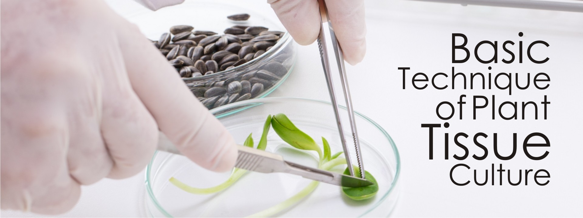
STERILIZATION OF EXPLANTS
Seeds, axillary and Apical meristem and first three axillary buds from nodal segments, leaves were collected from field grown matured plants. The explants were thoroughly washed with 2-4 drops of surfactant (Tween 20 or any soft liquid soap) for 10 minutes under running tap water. Surfactants reduce the surface tension of material being cleaned there by making the disinfectant solution more effective.
After washing with tap water properly the explants were subjected to chemical sterilization in the hood of laminar airflow with 0.1% w/v for 3-5 minutes. After HgC/l2 treatment the explants were washed 3-4 times with sterilized double distilled water to remove the traces of steril